Research

Graduate Student Ramya Vijayakumar First Author on Paper

Ramya Vijayakumar Circulation
Figure 1. Epicardial activation time (AT) isochrone maps. Examples of activation in ( left to right) control, Long QT Type 1 (LQT1) (patient 21), LQT2 (patient 3), and LQT3 (patient 7). In all LQTS types as in normal control, epicardial activation starts from breakthrough at anterior right ventricle (RV; shown by asterisk) near the RV outflow tract region, 20 to 30 ms after the onset of QRS. It proceeds in a uniform fashion to activate the ventricles synchronously. The latest region to activate is the left ventricle (LV) basal region (dark blue). The total ventricular activation time (TVAT) in all LQTS types was ≈50 ms, comparable with normal control. AO indicates aorta; LA, left atrium; ms, milliseconds; and RA, right atrium.

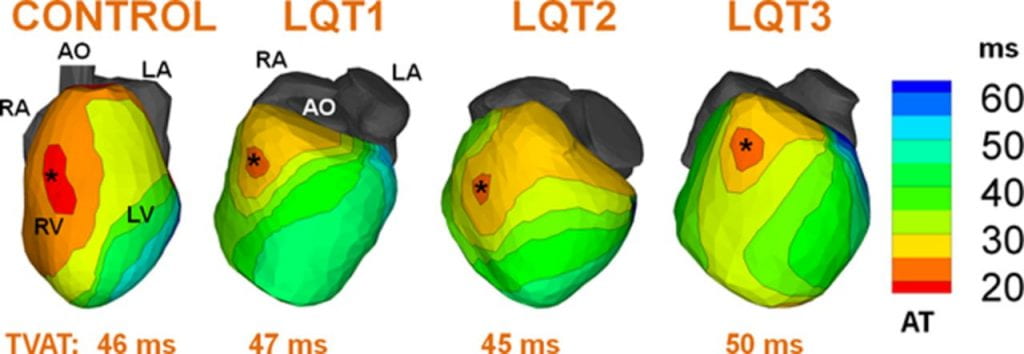
A recent publication in the journal Circulation entitled, “Electrophysiologic substrate in congenital Long QT syndrome: noninvasive mapping with electrocardiographic imaging (ECGI)” describes research conducted by Ramya Vijayakumar, a PhD student in the lab. Ramya used a novel noninvasive imaging modality – Electrocardiographic Imaging (ECGI) to study the arrhythmic substrate in the hearts of 25 patients with a hereditary rhythm disorder, called the Long QT syndrome, that can lead to sudden death. This is the first study to shed light on the abnormal electrophysiology of a genetic disorder in the intact human heart.

Leave a Reply

Your email address will not be published. Required fields are marked *